Endoptix: Endometriosis Diagnostics Device
Medical Device Engineering and Development
Medical Device Engineering and Development
Throughout the course of a single semester, my team developed a surgical aid device that uses narrow-band imaging and real-time image classification to help surgeons detect endometriotic lesions more accurately during minimally invasive procedures. By enhancing intraoperative visualization and reducing false negatives, the system aims to improve surgical outcomes and lower recurrence rates for patients with endometriosis.
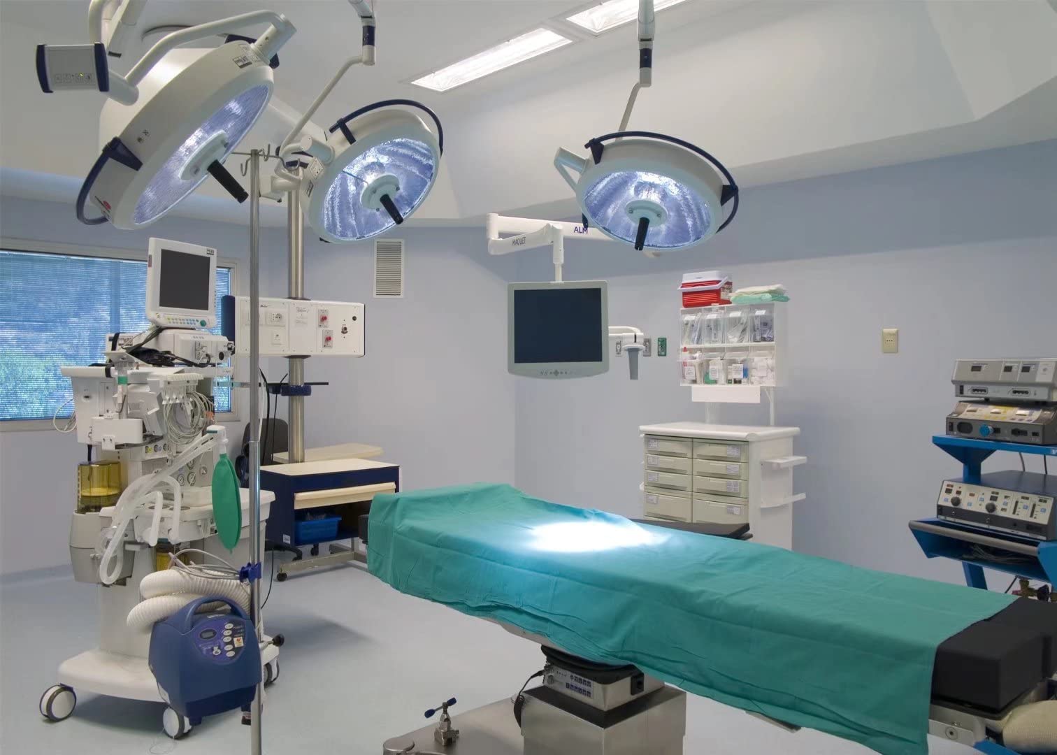
Endometriosis, a condition where uterine epithelial tissue grows outside the uterus, affects up to 10% of women of reproductive age and up to 50% of women with infertility. Despite its prevalence, patients often wait an average of seven years from symptom onset to treatment. Even after surgical intervention, recurrence rates can reach 50% within five years, often due to incomplete lesion removal. During minimally invasive laparoscopic surgery, surgeons must rely on visual identification to detect endometriotic lesions. Yet subtle or hidden lesions can be missed under standard white-light imaging. Current advanced optical technologies that might improve detection are often costly, bulky, and ill-suited for routine surgical use.
Our team designed a surgical aid device that combines novel narrow-band imaging (NBI) technology with custom image recognition and classification software to enhance intraoperative detection of endometriotic lesions. The device integrates a compact, flexible laparoscopic camera with a targeted blue-light source (~440 nm) to improve lesion contrast, paired with real-time image analysis to outline suspected tissue for the surgeon. This approach aims to improve diagnostic accuracy, reduce false negatives, and enable faster, more confident surgical decision-making—while maintaining an affordable, minimally invasive form factor compatible with existing laparoscopic workflows.
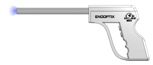
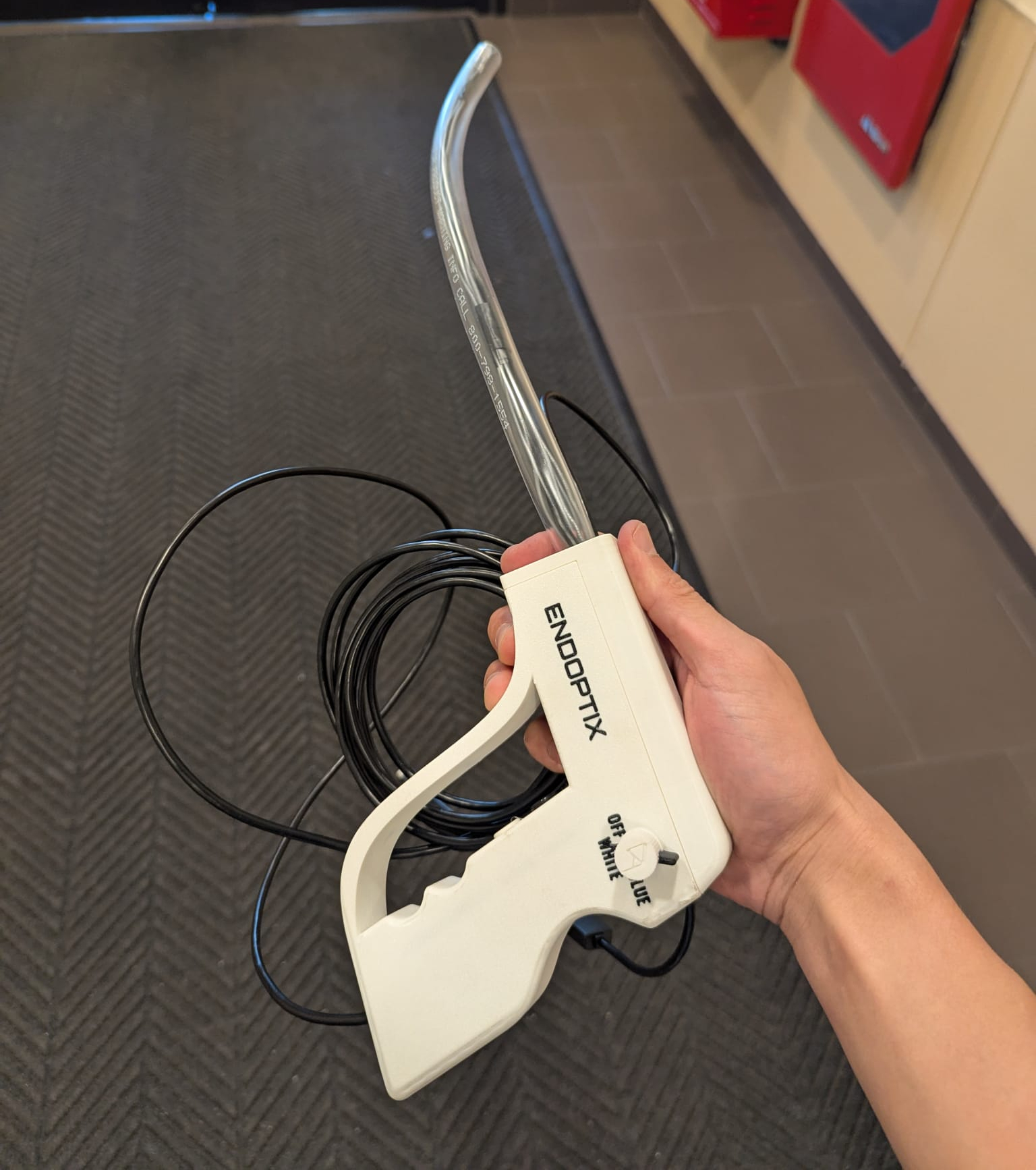
The design consists of a soft, elastomeric insertion tube housing both the light source and camera, connected to a 3D-printed handle and electronics enclosure containing a microcontroller, control circuitry, and activation button. Narrow-band blue light illuminates tissue in the Soret absorption band to enhance visualization of endometrial lesions, while high-resolution images are transmitted to a connected computer for spectral analysis. The surgeon receives real-time visual overlays highlighting areas flagged by the lesion detection algorithm. Key requirements included biocompatibility, safe and reliable operation inside a laparoscopic trocar, and a price point lower than current advanced optical imaging systems.
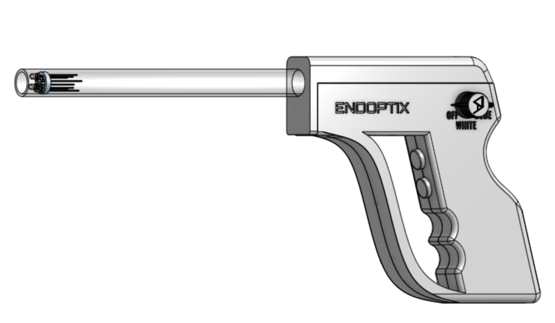
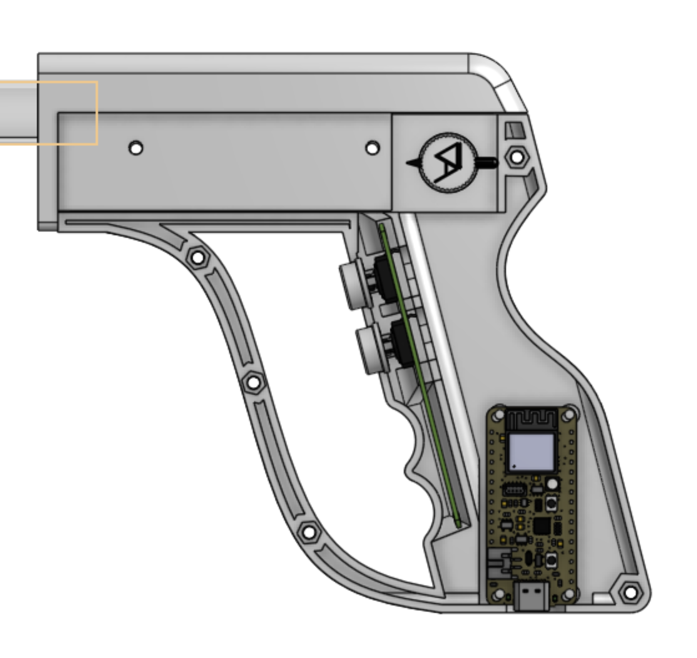
We fabricated a proof-of-concept prototype featuring micro-soldered high-intensity royal blue LEDs, precision-mounted alongside a 2 MP, 1920×1080 resolution laparoscopic camera. The elastomer tubing provided flexibility for navigation in minimally invasive procedures, while custom wire holders, snap-fit electronics housing, and a push-button trigger simplified surgeon interaction. The microcontroller handled image acquisition, data transfer, and initial processing before passing the feed to MATLAB for spectral analysis and lesion outlining. CAD modeling ensured the assembly met spatial constraints, and modular construction allowed for rapid component replacement during testing iterations.
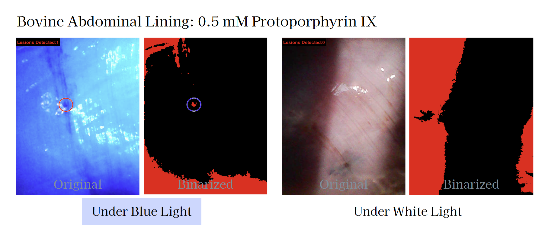
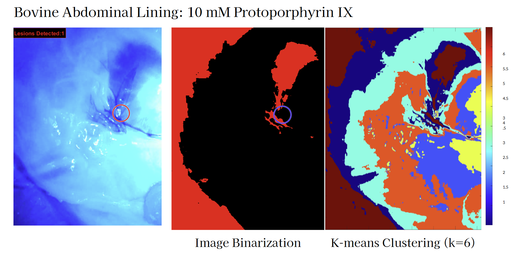
Two "killer experiments" guided feasibility testing. First, a murine uterus model with simulated endometriotic lesions (via Protoporphyrin IX injection) was imaged under both white light and narrow-band blue light to evaluate contrast enhancement. Second, a comparative study used annotated laparoscopic images to benchmark algorithm performance against expert and trainee identification, assessing sensitivity, specificity, precision, and accuracy. Early results confirmed that blue-light illumination improved lesion visibility in controlled conditions, and algorithm performance approached that of trained clinicians.
This project underscored the importance of validating both the hardware and software components under conditions that closely mimic the surgical environment. We found that optical enhancements like narrow-band imaging can offer measurable improvements in lesion visibility, but their impact depends heavily on precise wavelength control, adequate light intensity, and effective image processing algorithms. Iterative prototyping allowed us to refine the ergonomics, optical alignment, and data pipeline, while feedback from medical professionals highlighted usability factors that could influence adoption in the operating room. Additionally, benchmarking algorithm performance against expert annotations revealed that surgeon trust in such a device hinges not just on accuracy metrics, but on how seamlessly the system integrates into their workflow without adding complexity or time.